Genetics
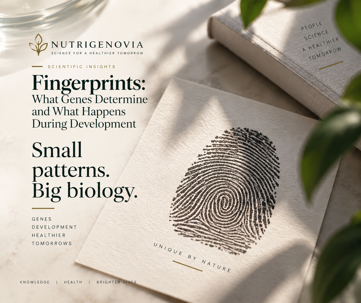
Fingerprints: What Genes Determine and What Happens During Development
- Author:
- NUTRIGENOVIA
- Published:
Fingerprints are one of those features we tend to think of as entirely individual. We use them to unlock our phones, confirm our identity, or simply leave them behind on the glass we are holding. Yet the fine ridges on our fingertips raise a biological question: how do they actually form?
Fingerprint ridges begin developing before birth. Genetics is involved, but the precise arrangement of every ridge is not stored in DNA as a finished set of instructions. It emerges while the fingers are growing and the skin covering them is developing.
Research into human development has now allowed scientists to follow this process much more closely. The familiar ridges on our fingertips arise through molecular signalling, changes in the shape of the developing finger, and interactions within the skin that can generate regularly spaced structures.
Fingerprints begin before birth
A developing hand is constantly changing in size and shape. During early fetal development, temporary swellings called volar pads appear on the fingertips, palms and soles. On the fingers, these pads are present during the same period in which the future fingerprint ridges begin to develop.
The timing matters because the ridges are forming on a surface that is itself changing. The fingertips grow, the volar pads regress, and the relationship between the epidermis and the tissue beneath it changes as development proceeds.
The primary ridges that will eventually produce the visible surface ridges begin forming during fetal development. By roughly the middle of pregnancy, the main fingerprint pattern has already been established. Later development adds further structural detail, but the basic organisation originates during this prenatal period.
Fingerprint development had been studied for decades before its molecular mechanisms were characterised in detail. Studies of human fetal skin described the formation of primary and secondary epidermal ridges and corresponding changes at the epidermal–dermal junction. Later theoretical work proposed mechanisms by which overall ridge patterns might arise. In 2005, Kücken and Newell developed a computational mechanical model in which differential growth, mechanical stresses and the changing geometry of the volar pads contributed to fingerprint formation.
A 2023 study published in Cell addressed the problem at the molecular and cellular level. James Glover and colleagues combined observations of human fetal tissue with experimental models and mathematical simulations. Their work provided experimental and modelling evidence implicating EDAR, WNT and BMP signalling and supported the involvement of a Turing reaction–diffusion system in ridge patterning.
How does skin start making ridges?
The earliest fingerprint ridges begin as small epithelial structures. At the molecular level, they resemble another familiar developmental structure: the early precursor of a hair follicle.
The resemblance is limited to this early epithelial stage. A developing hair follicle recruits a cluster of mesenchymal cells beneath the epithelium and continues through the programme required to produce a follicle. Fingerprint ridges do not recruit this mesenchymal condensate and do not proceed through the later stages of hair-follicle development. Instead, the epithelial structures extend into ridges.
Several signalling pathways are involved, including WNT, BMP and EDAR.
Mouse digit experiments provided the causal evidence: manipulating WNT, EDAR or BMP altered ridge formation and, depending on the perturbation, ridge spacing or thickness. Cultured human fetal volar skin showed pathway-responsive molecular and gene-expression changes. In human skin organoids, WNT increased epidermal proliferation and LEF1 expression, while BMP reduced proliferation; neither human system demonstrated the ridge phenotypes observed in mice.
The spacing between two fingerprint ridges therefore does not require a separate genetic instruction saying where each ridge should be placed. It can arise from molecular interactions taking place locally within developing tissue.
Alan Turing and the spacing between fingerprint ridges
In 1952, mathematician Alan Turing published a model describing how interacting substances moving through tissue could generate organised biological patterns.
Under the right conditions, an activating signal promotes a process locally while an opposing signal restricts it nearby. Repeated interactions between them can produce regularly spaced structures rather than uniform growth across the entire tissue.
This type of process is known as a reaction–diffusion system.
Mouse perturbation experiments support these pathway interactions, while mathematical modelling shows that interactions among EDAR, WNT and antagonistic BMP signalling can account for the regular spacing of the developing ridges.
Regular spacing alone, however, would not give us loops, arches and whorls.
The ridges do not begin forming everywhere on the fingertip at the same time. Ridge formation is initiated at several anatomically distinct sites, including areas associated with the centre of the volar pad, the dorsal–ventral boundary near the nail bed and the flexion crease. From these sites, waves of ridge formation spread across the developing fingertip. The timing and location of these waves affect how they meet.
When the researchers altered the initiation sites in their mathematical models, they could generate the major fingerprint pattern types: arches, loops and whorls.
The shape of a fingerprint therefore depends partly on a molecular pattern-forming system and partly on the geometry on which that system is operating.
Digit geometry, in turn, is influenced by genes active during limb development.
What do genes actually determine?
A large genome-wide study published in Cell in 2022 looked for genetic variants associated with fingerprint patterns.
Initial genome-wide analyses in Han Chinese cohorts identified 18 associated loci. A subsequent trans-ethnic meta-analysis identified 43 fingerprint-associated loci, and genes near those regions were strongly enriched for functions involved in limb development.
Some of the strongest genetic signals were not pointing primarily towards genes that build ridges in the skin. They were pointing towards genes involved in building the fingers themselves.
One of the variants investigated in detail lies near the MECOM locus, which encodes the EVI1 transcription factor. Experimental work in mice confirmed a role for EVI1 in dermatoglyph patterning, while its expression during human development suggested that it affects fingerprints through its role in shaping the limbs and digits rather than by directly controlling skin patterning.
Within two of the Han Chinese cohorts, comprising a combined 6,318 participants, the researchers also found genetic correlations between fingerprint patterns and hand proportions.
This gives us a rather different picture of what a “gene for fingerprints” would mean. A genetic variant does not need to tell skin cells exactly where to place a ridge. It can alter the development or proportions of the finger on which the ridge-forming system later operates.
Is your fingerprint hidden somewhere in your DNA?
Humans share the overwhelming majority of their DNA sequence. The fraction that varies between individuals contributes to an enormous range of biological differences, so it is tempting to imagine our individual traits as information that can simply be located somewhere among those genetic differences.
A fingerprint is not encoded in that way: no sequence of DNA contains a miniature map of every ridge ending, fork and curve on your index finger.
Genetic variation can influence fingerprint pattern type. Genes regulate the development of the fingers and the molecular machinery required for epidermal ridges to form. But the final configuration develops as those processes unfold in growing tissue.
Twins arising from the same fertilised egg begin with extremely similar inherited genomes. Their fingerprints can also resemble one another in broad pattern characteristics. They are not, however, identical in their fine ridge details.
The difference does not require a separate set of “fingerprint genes” in each twin. Each fetus undergoes its own development. The ridge-forming system operates on separate growing fingers, and small local differences during that process can contribute to different final configurations.
What happens when a genetic component is disrupted?
Rare cases in which normal epidermal ridges fail to form provide another line of evidence about the genetic contribution to fingerprints.
Adermatoglyphia is a rare condition characterised by the absence of normal epidermal ridges on the fingers, palms, toes and soles.
In 2011, researchers investigated a large family in which adermatoglyphia was inherited in an autosomal-dominant pattern. Their analysis identified a variant affecting SMARCAD1. More specifically, the variant disrupted a splice site associated with a short, skin-specific isoform of the gene. The variant segregated with adermatoglyphia throughout the family.
Further research identified additional SMARCAD1 variants affecting the same conserved splice site in other people with autosomal-dominant adermatoglyphia, strengthening the evidence that this skin-specific form of SMARCAD1 has an important role in dermatoglyph development.
SMARCAD1-associated isolated adermatoglyphia is one genetic route to absent dermatoglyphs, which can also occur as part of other inherited syndromes.
Cases like these put an important limit on any claim that fingerprints are simply the product of random events in the womb. Normal ridge development depends on genetic regulation. Disrupt part of that regulation, and the ridges themselves may fail to develop normally.
From DNA to a fingerprint
The formation of a fingerprint involves several biological levels at once. Genes influence the development and proportions of the fingers. Other genetic mechanisms are required for epidermal ridges to develop normally. WNT, BMP and EDAR signalling pathways help organise ridge formation within the skin. The shape of the growing fingertip provides the surface over which that process spreads. Ridge formation begins at particular anatomical locations, and the way those developing ridges meet contributes to the familiar arches, loops and whorls.
By the time the process is complete, the resulting ridge configuration is sufficiently individual for biometric identification.
The exact fingerprint was not stored intact in the genome; its detailed configuration emerged during development.
References
- Hale AR. Morphogenesis of volar skin in the human fetus. American Journal of Anatomy. 1952;91(1):147–181. doi:10.1002/aja.1000910105. DOI
- Penrose LS, O'Hara PT. The development of the epidermal ridges. Journal of Medical Genetics. 1973;10(3):201–208. doi:10.1136/jmg.10.3.201. DOI · PubMed
- Okajima M. Development of dermal ridges in the fetus. Journal of Medical Genetics. 1975;12(3):243–250. doi:10.1136/jmg.12.3.243. DOI · PMC
- Misumi Y, Akiyoshi T. Epidermal ridge formation in the human fetus: A correlation to the appearance of basal cell heterogeneity and the expression of epidermal growth factor receptor and cytokeratin polypeptides in the epidermis. American Journal of Anatomy. 1991;191(4):419–428. doi:10.1002/aja.1001910409. DOI · PubMed
- Kücken M, Newell AC. Fingerprint formation. Journal of Theoretical Biology. 2005;235(1):71–83. doi:10.1016/j.jtbi.2004.12.020. DOI · PubMed
- Glover JD, Sudderick ZR, Shih BBJ, et al. The developmental basis of fingerprint pattern formation and variation. Cell. 2023;186(5):940–956.e20. doi:10.1016/j.cell.2023.01.015. DOI · PubMed
- Turing AM. The chemical basis of morphogenesis. Philosophical Transactions of the Royal Society of London. Series B, Biological Sciences. 1952;237(641):37–72. doi:10.1098/rstb.1952.0012. DOI
- Li J, Glover JD, Zhang H, et al. Limb development genes underlie variation in human fingerprint patterns. Cell. 2022;185(1):95–112.e18. doi:10.1016/j.cell.2021.12.008. DOI · PMC
- Tao X, Chen X, Yang X, Tian J. Fingerprint recognition with identical twin fingerprints. PLoS ONE. 2012;7(4):e35704. doi:10.1371/journal.pone.0035704. DOI
- Jónsson H, Magnúsdóttir E, Eggertsson HP, et al. Differences between germline genomes of monozygotic twins. Nature Genetics. 2021;53(1):27–34. doi:10.1038/s41588-020-00755-1. DOI
- Nousbeck J, Burger B, Fuchs-Telem D, et al. A mutation in a skin-specific isoform of SMARCAD1 causes autosomal-dominant adermatoglyphia. American Journal of Human Genetics. 2011;89(2):302–307. doi:10.1016/j.ajhg.2011.07.004. DOI · PMC
- Nousbeck J, Sarig O, Magal L, et al. Mutations in SMARCAD1 cause autosomal dominant adermatoglyphia and perturb the expression of epidermal differentiation-associated genes. British Journal of Dermatology. 2014;171(6):1521–1524. doi:10.1111/bjd.13176. DOI
- Lugassy J, Itin P, Ishida-Yamamoto A, et al. Naegeli–Franceschetti–Jadassohn syndrome and dermatopathia pigmentosa reticularis: two allelic ectodermal dysplasias caused by dominant mutations in KRT14. American Journal of Human Genetics. 2006;79(4):724–730. doi:10.1086/507792. DOI · PMC